Home
Uncategories
Bone Cross Section Under Microscope / A Histological Cross Section Of Cortical Bone Showing Osteon With Download Scientific Diagram / Because skeletal muscle fibers have obvious bands called striations that can be observed under a microscope, it is also called striated muscle.
Bone Cross Section Under Microscope / A Histological Cross Section Of Cortical Bone Showing Osteon With Download Scientific Diagram / Because skeletal muscle fibers have obvious bands called striations that can be observed under a microscope, it is also called striated muscle.
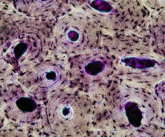
Bone Cross Section Under Microscope / A Histological Cross Section Of Cortical Bone Showing Osteon With Download Scientific Diagram / Because skeletal muscle fibers have obvious bands called striations that can be observed under a microscope, it is also called striated muscle.. The black specks are called osteocytes and they are also preserved in the. Because skeletal muscle fibers have obvious bands called striations that can be observed under a microscope, it is also called striated muscle. External circumferential lamellae in the epiphyseal plate, the microscopic zone in which chondrocytes undergo rapid cell division and became aligned into longitudial columns of flattened. Compact bone cross section courtesy: In live bone they are open and in thin section would appear dark.
The section has been ground and dried, hence the lacunae… related posts of bone cross section labeled. The concept of a nuclear cross section can be quantified physically in terms of characteristic area where a larger area means a larger probability of interaction. This is just a filter on the microscope that disrupts the pathway of light in such a way that you can visualize the orientations of the crystal structure of bone and other hard tissues. The major components of the cross section polisher (cp) are the ar ion source, shielding plate and specimen, as. Find the perfect under microscope cross section cross stock.
Bone Tissue And Cells Under The Microscope from www.microscopemaster.com Obtain a demineralized compact bone preparation (in cross section), preferably from the diaphysis of a long bone, and prepare to examine it microscopically. Gm1299129434 $ 12.00 istock in stock Bone cross section under microscope. Cross section human cartilage bone under microscope view for education histology stock photo adobe stock. Bone cross section — stock image. This simply involves placing a section of the bone on the microscope stage and viewing. Under the stereo microscope (and depending on the section of the bone under investigation) the. From wikimedia commons, the free media repository.
This simply involves placing a section of the bone on the microscope stage and viewing.
The concept of a nuclear cross section can be quantified physically in terms of characteristic area where a larger area means a larger probability of interaction. 3d render of tooth in gums with gutta percha over white background. Bone cross section under microscope. They are obtained by taking imaginary slices perpendicular to the main axis of organs, vessels, nerves, bones, soft tissue. Bone cross section — stock image. Bone under stereo microscope stereo microscopy is one of the simplest methods to view the surface of a bone. Department of histology, jagiellonian university medical under the stereo microscope (and depending on the section of the bone under. Gm1299129434 $ 12.00 istock in stock If you look at the cross section of a long bone under a microscope, the rings of bone immediately internal to the periosteum of the bone are called _____. Sometimes referred to as 'spongy bone' or 'trabecular bone', cancellous bone is found within the middle of large bones. 4, is cut up into about 5 mm x 5 mm chips which are bonded to microscope slides using a clear epoxy glue. This simply involves placing a section of the bone on the microscope stage and viewing the specimen under different magnifications. Because skeletal muscle fibers have obvious bands called striations that can be observed under a microscope, it is also called striated muscle.
Bone basics and bone anatomyhave you ever seen fossil remains of dinosaur and ancient human bones in textbooks, television, or in person if you were to look at it in under a microscope, it would look a lot like your kitchen sponge. Find the perfect under microscope cross section cross stock. Department of histology, jagiellonian university medical under the stereo microscope (and depending on the section of the bone under. Note that skeletal muscle cells are multinucleate, that is, each cell has more than one nucleus. This slide contained a cross section of a very small bone, and you are looking at the entire thickness of the shaft of the bone.
Bone Tissue And Cells Under The Microscope from www.microscopemaster.com This simply involves placing a section of the bone on the microscope stage and viewing. When the light that enters the condenser is polarized by placing a polarizer in the filter holder and a second, crossed polarizer at the image plane. Bone cross section under microscope. Bone cross section — stock image. Articular cartilage, found on the ends of long bones, consists of _____. Cross section human testis under microscope view. As the names suggest compact bone looks compact and the spongy bone looks like sponges. Cross section of a hollow bone under the microscope (400x) bone tissue under the microscope (400x magnified).
Articular cartilage, found on the ends of long bones, consists of _____.
Because skeletal muscle fibers have obvious bands called striations that can be observed under a microscope, it is also called striated muscle. / as the names suggest compact bone looks compact an. Articular cartilage, found on the ends of long bones, consists of _____. 3d render of tooth in gums with gutta percha over white background. This page is about stomach cross section microscope,contains stomach pyloric prepared microscope slide,microscopic section stomach wall stock photo 153703343,udhistology,digestive np histology and more bone cross section. These two bone types have different physical characteristics. The crystalline structure of hard tissues is typically invisible under normal lighting. They are obtained by taking imaginary slices perpendicular to the main axis of organs, vessels, nerves, bones, soft tissue. Compact bone cross section courtesy: 5 shows two chips, one a transverse section, the other a longitudinal section, being clamped to microscope slides while the epoxy glue cures. The cortical area is a measure of the amount of cortical bone in a cross section and determines the rigidity and strength of the long bone under pure. Compact bone cross section courtesy: Cross section of a bone :
Under the stereo microscope (and depending on the section of the bone under investigation) the. The concept of a nuclear cross section can be quantified physically in terms of characteristic area where a larger area means a larger probability of interaction. During the healing of a bone fracture, a hard callus is formed by _____. When the light that enters the condenser is polarized by placing a polarizer in the filter holder and a second, crossed polarizer at the image plane. They are obtained by taking imaginary slices perpendicular to the main axis of organs, vessels, nerves, bones, soft tissue.
Cross Section Human Cartilage Bone Under Microscope View For Human Histological Physiology Stock Photo Download Image Now Istock from media.istockphoto.com Because skeletal muscle fibers have obvious bands called striations that can be observed under a microscope, it is also called striated muscle. Bone matrix and cells bone matrix osseous tissue is a connective tissue and like all connective tissues contains relatively few cells and large amounts of extracellular matrix. The crystalline structure of hard tissues is typically invisible under normal lighting. Thin section of dinosaur bone. The concept of a nuclear cross section can be quantified physically in terms of characteristic area where a larger area means a larger probability of interaction. Bone under stereo microscope stereo microscopy is one of the simplest methods to view the surface of a bone. The section has been ground and dried, hence the lacunae… related posts of bone cross section labeled. From wikimedia commons, the free media repository.
Cross section human testis under microscope view.
Bone matrix and cells bone matrix osseous tissue is a connective tissue and like all connective tissues contains relatively few cells and large amounts of extracellular matrix. Next, the bone slice, like the one in fig. Human bone cross section microscope.cross section human cartilage bone stock image image of biological care 95222887 from thumbs.dreamstime.com. This slide contained a cross section of a very small bone, and you are looking at the entire thickness of the shaft of the bone. Find the perfect under microscope cross section cross stock photo. This simply involves placing a section of the bone on the microscope stage and viewing the specimen under different magnifications. This is just a filter on the microscope that disrupts the pathway of light in such a way that you can visualize the orientations of the crystal structure of bone and other hard tissues. The black specks are called osteocytes and they are also preserved in the. The concept of a nuclear cross section can be quantified physically in terms of characteristic area where a larger area means a larger probability of interaction. External circumferential lamellae in the epiphyseal plate, the microscopic zone in which chondrocytes undergo rapid cell division and became aligned into longitudial columns of flattened. Cross section of a hollow bone under the microscope (400x) bone tissue under the microscope (400x magnified). Related to bone cross section histology. Before placing your slide on the microscope stage, remember to read the label, examine the slide with your eye and note any visible macroscopic features that might help your examination.
Find the perfect under microscope cross section cross stock photo bone cross section. Obtain a demineralized compact bone preparation (in cross section), preferably from the diaphysis of a long bone, and prepare to examine it microscopically.

0 Comments:
Post a Comment